Interventional Radiology: Using 3-D Simulations to Enhance Radiation Protection of Medical Staff
In interventional radiology, diagnostic or therapeutic medical interventions can be performed while monitoring them at the same time by means of imaging. Such interventions increasingly offer an alternative to classical surgery as they usually do not require a general anaesthetic and are associated with fewer risks, less pain and shorter convalescence times. However, as ionising X-rays are often used for imaging, the radiation exposure of medical staff increases with the number of such operations. In an interdisciplinary research project funded by the Federal Ministry for the Environment, GRS scientists are developing a three-dimensional simulation model that will allow conclusions to be drawn on how to optimise radiation protection.
So-called interventional radiology can solve major health problems by relatively small interventions. Imaging procedures and special instruments are used to perform therapeutic or diagnostic procedures in a minimally invasive manner, i.e. without major surgery.
A concrete example of an application where interventional measures are used are narrowed coronary arteries, which can trigger coronary heart disease and even a heart attack. During treatment, the doctor inserts a very small balloon into the body using a guide wire and a catheter. The position of the balloon in the body can be followed live, so to speak, via selective x-rays. Once the balloon reaches the constricted area, it is inflated to reopen the vessel. Other illnesses where interventional measures are used include certain cancers and strokes.
Interventional radiology measures - in technical terms, this is referred to as "interventional cardiology" referring to the medical area in which the disease falls - often spare the patient classic surgery with a general anaesthetic and are usually associated with fewer risks, less pain and shorter convalescence times. Since the medical-therapeutic potential of these measures is constantly developing, image-guided interventions are now performed much more frequently than ten or twenty years ago.
Radiation protection law defines "limit values for occupationally exposed persons”
Due to the increasing number of such treatments, medical personnel are increasingly exposed to ionizing X-rays. In order to protect people who come into contact with ionizing radiation at work, German radiation protection law defines so-called "limit values for occupationally exposed persons". Since different parts of the body react differently to radiation, § 78 of the Radiation Protection Act stipulates different limit values (so-called organ dose equivalent) for different organs - such as the lens of the eye, the skin, or even the uterus in women capable of giving birth.
On the other hand, a limit value of 20 millisievert (mSv) per calendar year is prescribed for the effective dose, which already takes into account the different sensitivities of the organs. By way of comparison, a person flying from Munich to Tokyo is exposed to cosmic radiation of up to 0.1 mSv. The average annual radiation exposure of the German population from natural sources is 2-3 mSv.
A clear description of radiation exposure is difficult
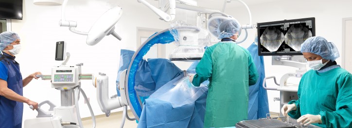
While the patients lie still during surgery and appropriate radiation protection measures (e.g. lead shielding) are applied, medical personnel move in the radiation field and are therefore not easily protected, especially regarding those parts of the body that are not covered by the obligatory lead apron. In order to measure the radiation exposure of personnel, so-called dosimeters are used, which are usually worn on the chest. As medical personnel move during operations, various parts of the body and organs are constantly exposed to X-rays. A measured value of the effective dose on the thorax can therefore not be assumed to be representative of the radiation exposure of the eye’s lens, for example. A further challenge is the large number of different interventional measures. Standardised times or sequences of movements on the basis of which the individual radiation exposure might be estimated are hardly possible.
In order to be able to make meaningful statements nevertheless, a GRS research team uses a three-dimensional simulation model developed in-house within the framework of a project funded by the Federal Ministry for the Environment to determine approximately the respective radiation exposure of the persons involved. From this model, conclusions can then be drawn to optimise radiation protection.
3-D simulation model developed in preliminary study
The scientists draw on a project also funded by the Federal Environment Ministry. In this project, a three-dimensional simulation model of the dose distribution in an intervention room was created using so-called Monte Carlo methods. Monte-Carlo methods use, in very simplified terms, statistical and probabilistic procedures to solve numerically mathematical problems that can only be solved with great effort or cannot be solved analytically at all.
Within the developed simulation model, the code package Geant4 can be used to simulate how particles interact with matter as they pass through it. Specifically, the research team simulated digitally a typical intervention room, mapping the dose distribution in the room caused by ionizing (scattered) radiation at different settings of the X-ray device. Using this 3D map, the dose can be calculated at each individual point in the room.
To validate the simulation results, the researchers, in cooperation with University Hospital Augsburg and University Hospital Cologne, collected real measuring data under defined, static conditions. For example, they measured the dose at different points in a real intervention room. This was done with the aid of dosimeters attached to "phantoms". The research team changed various parameters that also change during real interventional measures, such as the orientation of the X-ray machine.
Job exposure matrices for medical staff
The researchers are building on the results of this preliminary study in the follow-up study that is currently underway. One of the aims now is to create so-called job exposure matrices for medical personnel. With these matrices, it is possible to determine approximately to what levels of radiation the medical personnel concerned are exposed annually. For this purpose, anonymised data sets of real interventional measures are depicted in the simulation with the help of the relevant parameters contained therein (e.g. orientation of the X-ray machine, number of X-ray images taken during an intervention...), i.e. they are quasi-simulated. The researchers hope e.g. that it will be possible to estimate from the database obtained in this way how high a certain organ dose equivalent of a particular person in a calendar year will be.
Furthermore, concrete recommendations for radiation protection are to be derived from the results in order to keep the radiation exposure of the personnel as low as possible. This can be achieved on the one hand by using so-called radiation protection equipment. This may comprise e.g. lead curtains under the patient tabletop or movable acrylic lead glass panels mounted on the ceiling that shield the staff from radiation. With the aid of simulation, it is possible to investigate systematically at which position a piece of radiation protection equipment will give optimum protection. Also, recommendations can be derived as to the positions in the room where personnel are exposed to the lowest possible level of radiation.
The project, which is also funded by the BMU, has a term of three years and will run until the end of March 2023.
Sven Dokter
GRS gGmbH
sven.dokter@grs.de